
The structure of the ankle joint is a sophisticated interplay of stiffness and mobility. The mobility of the ankle joint is limited by ligaments and tendons. However, these provide the ankle with its stability and functionality. Below, we will take a closer look at the anatomy of the ankle joint and show you the general structure of the ankle joint and its most important ligaments.
Ankle anatomyThe ankle joint is the first joint in the human body, seen from the foot to the head. Compared to the upper joints of the extremities, such as the shoulder or wrist, the ankle joint has very limited mobility. Its primary function is static. The ankle joint is roughly divided into an upper ankle joint (talocrural joint) and a lower ankle joint (talotarsal joint). Together, these two joints form a functional cylindrical joint (cylindrical joint).


Knowledge of the structure of the ankle joint is crucial for understanding the stability and support function of the foot. The main movements of the foot result from the functional interaction of the upper and lower ankle joints. The function of the upper ankle joint is extension and flexion (bending and stretching) of the foot. This function unfolds its effect in the dynamic locomotion of the body. The lower ankle joint is somewhat more complex in its structure and mechanical function.
The lower ankle and foot are capable of rotating movements around the vertical axis of the lower leg, inwards (adduction) and outwards (abduction). These rotations are known as supination (inward rotation of the sole of the foot) and pronation (outward rotation of the sole of the foot). This ensures the best possible contact surface on uneven surfaces and maximizes the stability of the ankle and foot. Each person has a different level of flexibility when it comes to maximum ankle movement. This mostly depends on age, the training status of the foot, and whether there has been damage to the structures involved in the past. The separate movements of the upper and lower ankle joints are limited. Only the sum of the two partial joints results in a greater range of motion, in which both complement each other to form a functional unit. The reason for this is the ligament and tendon systems that both joints share.
Ankle ligamentsThe structure and composition of the individual parts of the upper and lower ankle joints form the passive part of the movement chain. Movement of the joints is only possible through their connection to the muscles, ligaments, and tendons.

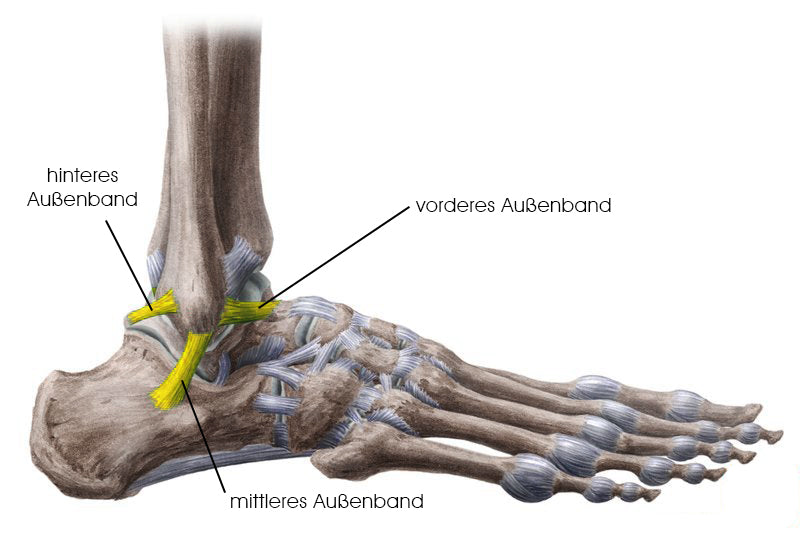
On the outside of the ankle are arguably the most vulnerable ligaments of the foot, the lateral ligaments. They limit the supination movement (turning the sole of the foot inward) in the lower ankle joint. In a so-called supination injury ("twisting"), the lateral ligaments of the ankle are often torn or overstretched. The lateral ligaments of the ankle are divided into three categories according to their location and function: the anterior, middle, and posterior lateral ligaments. All three attach to the lateral malleolus (outer malleolus). The lateral malleolus is the lower, thickened end of the fibula.
Ankle and Achilles tendonThe Achilles tendon is the strongest tendon in the human body. Its insertion is located on the entire surface of the calcaneal tuberosity. From there, it runs upwards towards the triceps brachii muscle. Its extreme tensile strength enables maximum power transmission from the calf muscles when extending the foot. It is often exposed to extreme situations in athletic activities. For example, in an 8-meter long jump, forces of up to 14,700 Newtons (N) are exerted, which corresponds to a tensile force of 1,500 kilograms (kg). The tensile strength of the Achilles tendon is far greater under dynamic loads than under static loads. One experiment demonstrated a maximum static tensile strength of 680 kg and a maximum dynamic tensile strength of 930 kg. Thus, the Achilles tendon is far more stable under short dynamic loads than under static loads. This property is extremely important and valuable for athletic use.